
For decades in the life-cyle of a person, our proteins will behave as expected. But at some point, some of those proteins will abandon their normal functions and become amyloid fibrils. When first discovered, amyloid fibrils were synonymous with diseases like Alzheimer’s Disease, ALS, Parkinson’s Disease, prion diseases, and others.
…But then amyloid was discovered in bacterial homes called biofilms, in mussels that use amyloid as glue to stick to rocks, and even in our own bodies that use amyloid to make skin pigment.
As scientists realized their abundance in nature, the idea that amyloid was more than just a disease-related issue began to take hold.
My current research seeks to understand why particular proteins that reversibly aggregate in our cells are also conspicuously found irreversibly aggregated in amyloid diseases.
I work with amyloid in all forms: the good and the bad.
The Water story
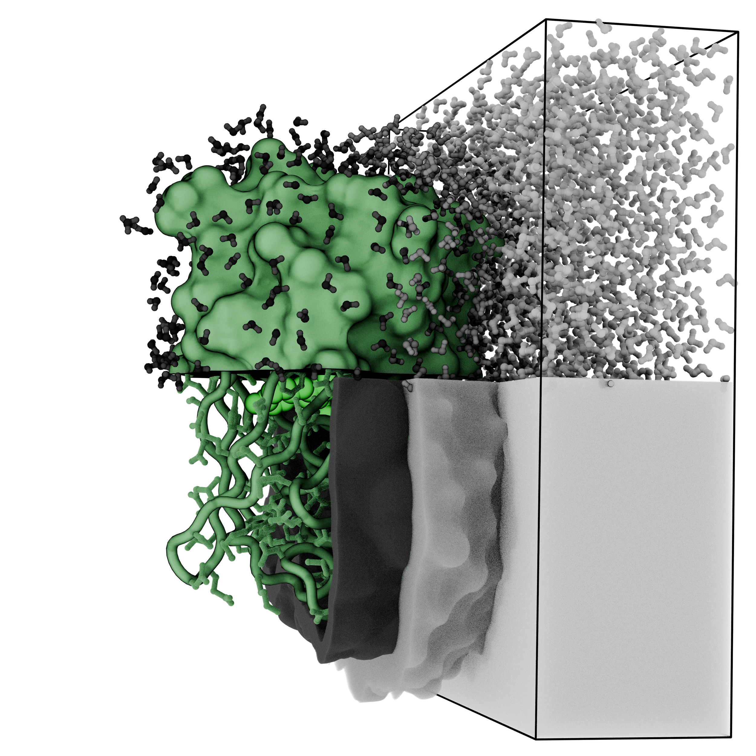
The PhD work that I did on LARKS and bridging protein assembly between amyloid and Liquid-Liquid Phase Separation (LLPS) led me to be interested in how the aqueous environment of the dense and light phase were different. As it turns out, protein motions and water motions are different in each aqueous phases of an LLPS system. This could have biological implications, if certain parts of a protein expand or contract upon entering into different aqueous phases in the cell, or if particular enzymatic chemistry is favored in one phase or another.
Re-examining cell biology with the lens of the cytoplasm, nucleoplasm, and membraneless organelles all behaving as distinct LLPS phases leads to so interesting insights in understanding how the cell is regulated as a biophysical system. Changes in gene expression, cell fate, and hydration can all be interrelated through understanding the underlying aqueous phase. Molecules will tend to enrich into a preferred aqueous phase as a result of balancing electric and osmotic potentials between the phases. This grand story is detailed in a 2025 open access review: Rethinking Cellular Organization: Phase Separation as a Unifying Principle in Molecular Biology.
The LARKS story
In 2012, some RNA-binding proteins were found to form hydrogels that were composed of amyloid-like fibrils.
These hydrogels would melt back into a liquid when heated, but then would reform into the amyloid fibrils that compose the hydrogel—a strikingly unique behavior because the doctrine was that amyloid states were considered ultrastable, i.e., not prone to melting.
For my doctoral thesis, I studied these proteins and discovered the fibrils they made had sharp kinks in the peptide backbone. A consequence of this kink is that it breaks up the surface area available for fibrils to interact with each other, which leads to less stable fibrils.
I called these structures Low-complexity Amyloid-like, Reversible Kinked segments or “LARKS.”

Figure examples
It can be hard to translate the richness of data available in 3D structures into 2D stories. Some examples of my best attempts are below.

Amyloid fibrils can be hard to depict. The cylinders give a sense of the space while also communicating strong vertical lines to orient the viewer. Only depicting the top layer improves clarity, and because most fibrils are parallel, in-register strands, every strand is close to identical to the ones above and below it. We only need to see one layer for most applications.
These kinds of representations of amyloid fibrils were pioneered by Dr. Michael Sawaya. Check out his Amyloid Atlas here.

Above is a representation of hnRNPA2 determined by cryoEM as determined in Lu et al., 2020. PDBID 6WQK. Below is the structure of luciferase colored by a propensity to form steric zippers as judged by zipperDB.
